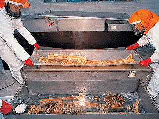

Decay is a big obstacle to the study of anatomy, so scientists have been searching for centuries for suitable preservation techniques. With the invention of plastination, it has become possible to preserve decomposable specimens in a durable and lifelike manner for instructional, research and demonstration purposes. During a vacuum process, biological specimens are penetrated with a reactive polymer developed specifically for this technique. The class of polymer used determines the mechanical (flexible or hard) and optical (transparent or opaque) properties of the preserved specimen. Plastinated specimens are dry and odorless; they retain their natural surface relief and are identical with their state prior to preservation down to the microscopic level. Even microscopic examinations are still possible. The plastination technique replaces bodily fluids and fat with reactive polymers, such as silicone rubber, epoxy resins, or polyester. In a first phase, solvent gradually replaces bodily fluids in a cold solvent bath (freeze substitution). After dehydration, the specimen is put in a solvent bath at room temperature to dissolve and remove the fat. The dehydrated and defatted specimen is then placed into a polymer solution. The solvent is then brought to a boil in a vacuum and continuously extracted from the specimen. The evaporating solvent creates a volume deficit within the specimen, drawing the polymer gradually into the tissue. After the process of forced impregnation, the specimen is cured with gas, light, or heat, depending on the type of polymer used.
“Slice plastination” is a special variation of this preservation technique. When applying this method, whole bodies or body parts (mostly deep-frozen) are first cut or sawed into 2-8 mm thick slices. These slices are then placed between wire nettings, where they are dehydrated, defatted and finally saturated with polymers in a vacuum. The impregnated slices are cured between sheets of film or cast with additional polymers in a flat chamber composed of glass plates to give them a smooth surface. The refraction index of the applied resins determines the optical properties of plastinated body slices. Body and organ slices produced with epoxy resins result in transparent specimens with good coloration of individual tissues. Polyester resins permit an excellent distinction between white and grey brain matter and are thus used for the plastination of brain slices. Plastinated organs and body slices are a novel teaching aid for cross-sectional anatomy, which is gradually gaining importance and can be easily correlated with radiological imaging. Series of transparent body slices are helpful for a large variety of scientific research activities. In addition, they are a suitable diagnostic means in pathology, as they allow rapid macroscopic and diagnostic screening of entire organs or operation preparations. Additionally, they still allow for selective analyses of pathological tissue regions with conventional microscopic methods.
Gunther von Hagens invented plastination at the Institute for Anatomy at Heidelberg University in 1977, and has developed it further ever since. Plastination has gained general acceptance and is carried out in many institutions throughout the world. The durability and lifelike state of plastinated specimens as well as their high instructional value have contributed to this acceptance. For more information about plastination, visit the BODY WORLDS website
No comments:
Post a Comment